
Why the link matters
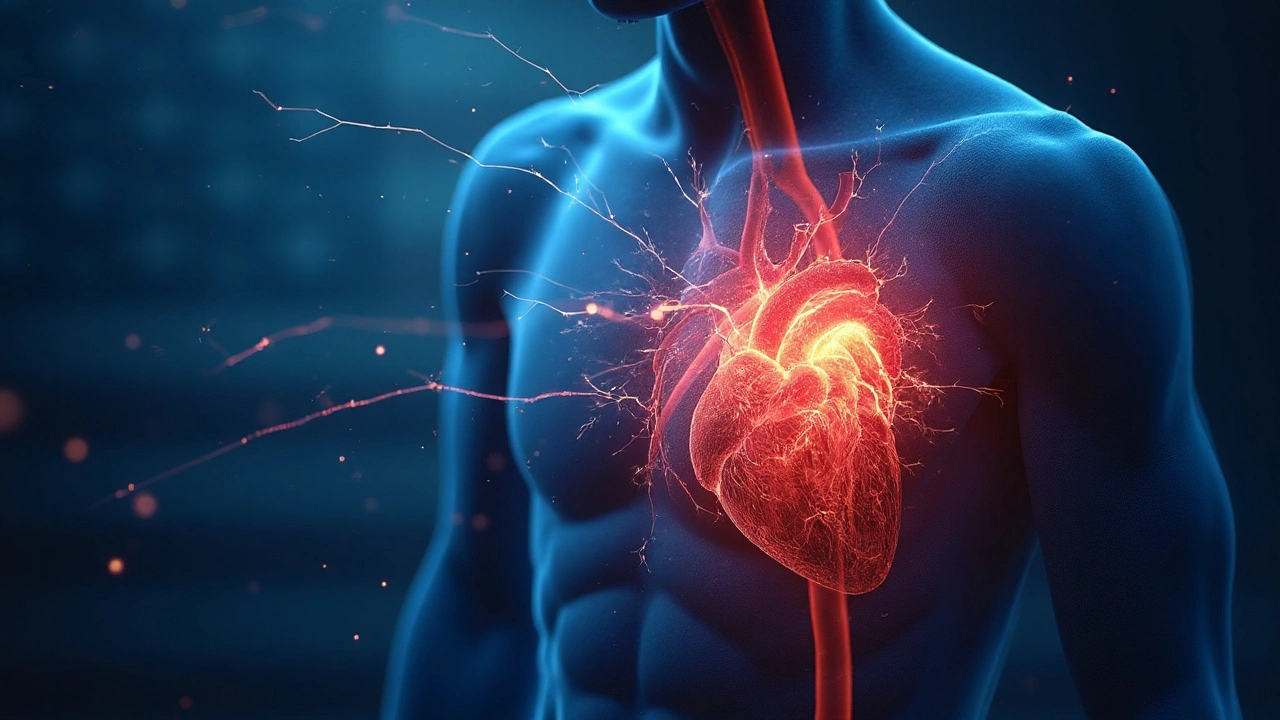
When the heart’s rhythm goes off‑beat, the left ventricle often pays the price. Understanding arrhythmias and left ventricular failure helps clinicians spot trouble early and choose therapies that keep the pump working.
Arrhythmia is a disturbance in the heart’s electrical timing that can be too fast, too slow, or irregular.Common forms-atrial fibrillation, ventricular tachycardia, and premature beats-alter how blood moves through the chambers. Over time, this can strain the left ventricle, the main pumping chamber, leading to Left Ventricular Failure, a subtype of heart failure where the ventricle cannot eject blood efficiently.
Pathophysiology: From rhythm glitches to pump breakdown
Three inter‑linked mechanisms explain the connection:
- Hemodynamic overload - Rapid rates shorten diastole, cutting coronary perfusion and raising wall stress.
- Neurohormonal activation - Irregular beats trigger the renin‑angiotensin‑aldosterone system (RAAS) and sympathetic surge, which promote fibrosis.
- Cardiac remodeling - Prolonged strain reshapes the ventricle, reducing Ejection Fraction, a key measure of contractility.
These processes feed each other, creating a vicious cycle where the arrhythmia worsens LV function, and the failing ventricle fuels more electrical instability.
Major arrhythmias that tip the balance
Not all irregular beats are equal. Below, the most prevalent culprits are compared.
| Arrhythmia | Typical Rate (bpm) | Key Hemodynamic Effect | Long‑Term LV Impact |
|---|---|---|---|
| Atrial Fibrillation | 100‑180 | Loss of atrial kick, irregular ventricular response | Reduced EF, dilated LV, higher mortality |
| Ventricular Tachycardia | 150‑250 | Severe reduction in stroke volume, acute ischemia | Rapid progression to pump failure, sudden cardiac death risk |
| Premature Ventricular Contractions (PVCs) | Variable | Irregular filling, post‑extrasystolic pause | In high burden (>10,000/day) may cause LV dysfunction |
Clinical assessment: Spotting the danger early
Doctors combine history, ECG, and imaging. Key steps:
- Identify symptom pattern - palpitations, exertional dyspnea, or syncope.
- Perform a 12‑lead Electrocardiogram to capture rhythm type and rate.
- Order a transthoracic echocardiogram to measure Ejection Fraction and look for LV dilation.
- Consider cardiac MRI when fibrosis is suspected - it predicts arrhythmia recurrence.
Blood biomarkers (BNP, NT‑proBNP) help gauge severity, while Holter monitoring quantifies PVC burden.
Treatment strategies that break the cycle
Therapy aims at two fronts: correcting the rhythm and supporting the ventricle.
Rate and rhythm control
Beta‑blockers (e.g., carvedilol) blunt sympathetic drive, lower heart rate, and improve survival. ACE inhibitors or ARBs reduce afterload, slowing remodeling.
For atrial fibrillation, options include:
- Pharmacologic cardioversion with flecainide or amiodarone.
- Electrical cardioversion for rapid restoration of sinus rhythm.
- Catheter ablation - shown in trials to lower HF hospitalizations.
Ventricular tachycardia often mandates an Implantable Cardioverter‑Defibrillator (ICD) to abort life‑threatening runs, plus anti‑arrhythmic drugs (e.g., sotalol) when needed.
Optimizing left ventricular function
Guideline‑directed medical therapy (GDMT) remains cornerstone:
- ARNI (sacubitril/valsartan) - superior EF improvement vs ACEI.
- Mineralocorticoid receptor antagonists - reduce fibrosis.
- Loop diuretics for congestion control.
When EF ≤35% despite GDMT, cardiac resynchronisation therapy (CRT) can re‑coordinate ventricular contraction, lowering arrhythmia burden.
Prevention and future directions
Primary prevention focuses on modifiable risk factors: hypertension, obesity, sleep apnea, and excessive alcohol. Early rhythm monitoring in at‑risk patients (e.g., post‑MI) catches silent PVCs before they overwhelm the LV.
Emerging therapies include:
- Gene‑based modulation of ion channels (still experimental).
- Newer sodium‑glucose cotransporter‑2 (SGLT2) inhibitors - they improve LV remodeling and cut arrhythmic events.
- Artificial‑intelligence‑driven ECG analysis for pre‑emptive identification of high‑risk patterns.
Related concepts and next steps
Understanding this connection sits inside a broader heart‑failure knowledge cluster. The next logical reads are:
- Neurohormonal activation in chronic heart failure
- Guideline‑directed medical therapy for reduced EF
- Device therapy: ICD vs CRT - when to choose
Each of these topics expands on a piece of the puzzle, helping clinicians craft a full‑spectrum care plan.
Frequently Asked Questions
Can a single episode of atrial fibrillation cause left ventricular failure?
A one‑off AF episode usually won’t produce permanent LV dysfunction, but repeated or sustained AF can erode atrial contribution to filling, raise filling pressures, and eventually reduce ejection fraction if untreated.
What PVC burden is considered dangerous for the left ventricle?
Studies show a daily PVC count above 10,000 (or >10% of total beats) is linked to measurable LV dilation and a drop in EF. Reducing the burden with ablation often improves function.
When should an ICD be implanted in a patient with ventricular tachycardia and LV failure?
Guidelines recommend ICD placement for secondary prevention in anyone who survived a sustained VT or VF, and for primary prevention when EF ≤35% despite optimal medical therapy, regardless of symptom burden.
Do beta‑blockers help both arrhythmia control and LV function?
Yes. Beta‑blockers slow heart rate, lessen sympathetic triggers for ectopy, and lower myocardial oxygen demand, which together improve EF and reduce hospitalization risk.
Is catheter ablation curative for atrial fibrillation‑related LV failure?
Ablation can restore sinus rhythm and halt further LV deterioration, but “cure” depends on how long AF persisted and the extent of existing remodeling. Early referral yields the best functional recovery.
mark Lapardin
Arrhythmias certainly remodel the myocardium, and the cascade of neurohormonal activation you described is spot‑on. The loss of atrial contribution during AF really spikes left‑ventricular filling pressures, nudging the ventricle toward eccentric hypertrophy. From a hemodynamic standpoint, tachyarrhythmias truncate diastole, which limits coronary perfusion and fuels ischemic insults. Overall, the interplay you outlined underscores why early rhythm control matters in preserving LV function.
September 25, 2025 AT 00:10
Barry Singleton
The piece glosses over the fact that not every PVC load is pathological; a threshold really matters. Also, the table could benefit from quantitative data on stroke volume reduction. Still, the mechanistic links are well‑summarized.
September 26, 2025 AT 03:57
Javier Garcia
Beta‑blockers blunt sympathetic drive and improve EF.
September 27, 2025 AT 07:43
christian quituisaca
Great synthesis! You’ve captured the vicious cycle between electrical instability and structural remodeling beautifully. The way you broke down hemodynamic overload, neurohormonal surge, and remodeling into three clear steps makes it easy to digest. I especially appreciate the emphasis on early detection with Holter monitoring – catching a high PVC burden before it remodels can be a game‑changer. Keep spreading this knowledge; it helps clinicians act before the pump breaks down.
September 28, 2025 AT 11:30
Donnella Creppel
Whoa!!! This article kinda skim‑s the surface – but let’s be real, not every arrhythmia is a death‑sentence!!! The author should have dived deeper into the molecular pathways – like, where’s the talk about oxidative stress??!! Also, the table layout could use more flair – maybe some color‑coding??!!
September 29, 2025 AT 15:17
Jarod Wooden
The relationship between arrhythmic burden and left ventricular failure is not merely a linear correlation; it is a profound illustration of how electrical disarray begets mechanical incompetence. When the myocardium is forced to fire at rates that eclipse its intrinsic refractory periods, diastolic filling time contracts, depriving the subendocardial fibers of essential perfusion. This iatrogenic ischemia leads to myocyte apoptosis, which cumulatively manifests as fibrosis – a substrate that predisposes to further arrhythmias. Moreover, the persistent activation of the renin‑angiotensin‑aldosterone system and sympathetic outflow creates a maladaptive feedback loop, intensifying afterload and promoting adverse remodeling. In the context of atrial fibrillation, the loss of atrial kick reduces preload, which, combined with irregular ventricular response, precipitates a precipitous drop in stroke volume. Ventricular tachycardia, by virtue of its high rates, can acutely diminish cardiac output to the point of hemodynamic collapse, heralding sudden cardiac death if not promptly terminated. Premature ventricular contractions, when frequent, generate post‑extrasystolic pauses that further destabilize ventricular filling dynamics, a phenomenon often underestimated in clinical practice. The thresholds cited – such as a PVC burden exceeding 10,000 per day – are not arbitrary; they reflect a tipping point where the heart’s compensatory mechanisms are overwhelmed. Therapeutically, this underscores the necessity of early rhythm control strategies, ranging from pharmacologic suppression to catheter ablation, especially in patients with borderline ejection fractions. Additionally, guideline‑directed medical therapy, including ARNIs and mineralocorticoid receptor antagonists, serves to blunt the neurohormonal cascade, thereby mitigating fibrosis and remodeling. Finally, device therapy, whether an ICD for secondary prevention or CRT for dyssynchrony correction, should be considered in the context of persistent arrhythmia‑induced LV dysfunction. In sum, arrhythmias are both cause and consequence of left ventricular failure, forming a self‑reinforcing loop that demands a multifaceted therapeutic approach.
September 30, 2025 AT 19:03
lee charlie
Seeing the link laid out like this really helps demystify why patients feel worse during fast rhythms. The emphasis on beta‑blockers and GDMT feels reassuring. Early detection truly can spare a lot of heart damage.
October 1, 2025 AT 22:50
Greg DiMedio
Oh great, another “must‑do” therapy list. Like we didn’t already have enough meds to juggle.
October 3, 2025 AT 02:37
Badal Patel
Indeed, one must contemplate whether the grandiose “future directions” are not merely a mirage of academic optimism; however, the inclusion of AI‑driven ECG analysis does provide a modicum of tangible advancement! In any case, let us not discard the allure of gene‑based modulation, even if it remains nascent. Lastly, the reverence for SGLT2 inhibitors is well‑placed, albeit perhaps overstated.
October 4, 2025 AT 06:23
KIRAN nadarla
The article could improve its citation density; many statements lack references. Also, “overload” should be “overload,” not “overload.” Minor but important.
October 5, 2025 AT 10:10
Kara Guilbert
Honestly, promoting PVC ablation without robust evidence feels ethically questionable. We mustn't rush into invasive procedures. Patient safety should be paramount.
October 6, 2025 AT 13:57
Sonia Michelle
Philosophically, the interplay between rhythm and pump illustrates the heart as both conductor and instrument. Practically, this means we should synchronize our therapeutic modalities, aligning rhythm control with ventricular support. The article’s structured approach mirrors this harmony. It also reminds us that evidence‑based medicine and compassionate care are not mutually exclusive. Ultimately, our goal remains to restore both electrical and mechanical integrity for the patient.
October 7, 2025 AT 17:43
Neil Collette
Well, if you think the author knows everything, you’re in for a disappointment. The “emerging therapies” section is pure hype without data. Readers deserve more than buzzwords.
October 8, 2025 AT 21:30
James Lee
Honestly the piece feels like it’s trying too hard to sound profound. A bit of brevity would have helped.
October 10, 2025 AT 01:17
Dennis Scholing
Thank you for compiling these pathways so clearly. The delineation between rate control and ventricular support is especially useful. Moreover, highlighting CRT as a bridge therapy adds practical insight. I look forward to future updates on real‑world outcomes.
October 11, 2025 AT 05:03
Kasey Lauren
This is a solid overview. It makes a complex topic approachable.
October 12, 2025 AT 08:50
joshua Dangerfield
I think the article nails the main points, but could add a bit on lifestyle tweaks like diet and stress. Also, a quick guide for primary care docs would be handy. Overall, great job.
October 13, 2025 AT 12:37
Abhimanyu Singh Rathore
Excellent breakdown! The mention of AI‑based ECG analysis is exciting-could revolutionize early detection!!! Also, the discussion on SGLT2 inhibitors is spot‑on; they truly offer cardio‑protective benefits. One minor note: “renin‑angiotensin‑aldosterone” should be hyphenated consistently.
October 14, 2025 AT 16:23
Stephen Lewis
Your summary adeptly bridges pathophysiology with clinical application. Emphasizing early rhythm monitoring aligns well with current guidelines. The inclusion of device therapy nuances adds depth. I appreciate the balanced tone throughout.
October 15, 2025 AT 20:10
janvi patel
While the article is thorough, I remain skeptical about the universal applicability of ablation in all PVC‑heavy patients. More stratified data would be beneficial.
October 16, 2025 AT 23:57